- 產品描述
美國CORTEZ進駐非洲瘧疾檢測試紙
廣州健侖生物科技有限公司
(廣州健侖生物科技有限公司是集研制開發、銷售、服務于一體的優良企業,公司產品涉及臨床快速診斷試劑、食品安全檢測試劑,違禁品快速檢測,動物疾病防疫檢測試劑,免疫診斷試劑、臨床血液學和體液學檢驗試劑、微生物檢驗試劑、分子生物學檢驗試劑、臨床生化試劑、有機試劑等眾多領域,同時核心代理Panbio、FOCUS、Qiagen、IBL、CORTEZ、Fuller、Inbios、BinaxNOW、LumuQuick、日本富士、日本生研等多家有名診斷產品集團公司產品,致力于為商檢單位、疾病預防控制中心、海關出入境檢疫局、衛生防疫單位,緝毒系統,戒毒中心,檢驗檢疫單位、生化企業、科研院所、醫療機構等機構與行業提供*、高品質的產品服務。此外,本公司還開展食品、衛生、環境、藥品等多方面的第三方檢測服務。)
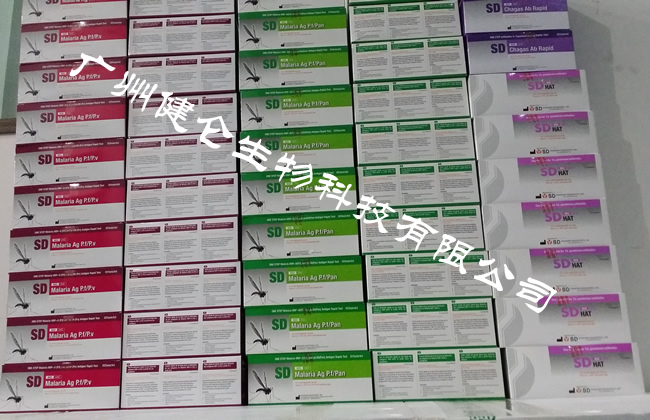
美國CORTEZ進駐非洲瘧疾檢測試紙 本試劑盒主要是采用膠體金層析的原理制成,用于檢測人體血清/血漿/全血標本中,感染的瘧原蟲抗體,包括了惡性瘧原蟲和間日瘧原蟲、卵形瘧原蟲、三日瘧原蟲共有抗原的鑒別性檢測。
人群易感性 人群對瘧疾普遍易感,感染后雖有一定的免疫力,但不持久,各型瘧疾之間亦無交叉免疫性,經反復多次感染后,再感染時癥狀可較輕,甚至無癥狀,而一般非流行區來的外來人員常較易感染,且癥狀較重。
People susceptible to the crowd generally susceptible to malaria, although the infection after a certain degree of immunity, but not lasting, there is no cross-immunity between malaria, after repeated infections, re-infection symptoms may be lighter, or even Asymptomatic, while the non-endemic areas of non-migrant workers are often more susceptible to infection, and the symptoms are severe.
:
1 撕開檢測卡鋁箔袋,取出袋內金標卡。注意:不要讓袋內材料暴露于高溫高濕環境,撕開鋁箔袋后盡快使用。
2將金標卡平放在臺面上;并將病人名字和編號寫在標簽上。
3 取5微升(吸管*刻度處)全血標本,垂直加入金標卡上“加樣孔A”內。
4 掰斷裂解液瓶子蓋子上方的綠色圓頭,在“樣品孔B”上垂直滴加4滴裂解液。
5 在十五分鐘內出結果。注意:必須在15分鐘內判讀結果,如超時判斷,結果無效。
6 請遵循相關法規,妥善處理樣本及廢棄材料。
7 存儲條件:2-30℃;
8 保質期:18個月;
【病原學檢測】
瘧疾檢測,用于檢測出虐疾的病原體——瘧原蟲,是明確診斷的zui直接證據。目前常用的層析法,具有操作簡單、靈敏度高和可鑒別蟲種等優點,廣泛用于瘧疾的病原學診斷,是目前zui常用的方法之一。
我司為美國NOVABIOS公司在中國地區戰略合作伙伴,負責該公司產品的總經銷及售后服務工作。還與各疾控中心,疾病防御中心有合作關系,例如中國疾病預防控制中心 、浙江省疾病預防控制中心 ,詳情可以我司工作人員。
( MOB:楊永漢)
我司還提供其它進口或國產試劑盒:登革熱、瘧疾、流感、A鏈球菌、合胞病毒、腮病毒、乙腦、寨卡、黃熱病、基孔肯雅熱、克錐蟲病、違禁品濫用、肺炎球菌、軍團菌、化妝品檢測、食品安全檢測等試劑盒以及日本生研細菌分型診斷血清、德國SiFin診斷血清、丹麥SSI診斷血清等產品。
廣州健侖生物長期供應各種違禁品檢測試紙、違禁品檢測卡、違禁品檢測試劑盒、藥篩試紙、藥篩試劑盒、嗎啡檢測試劑盒、巴比妥檢測試劑盒等。
想了解更多的產品及服務請掃描下方二維碼:

【公司名稱】 廣州健侖生物科技有限公司
【市場部】 楊永漢
【】
【騰訊 】
【公司地址】 廣州清華科技園創新基地番禺石樓鎮創啟路63號二期2幢101-103


隨著學科的發展,病理學的研究手段已遠遠超越了傳統的經典的形態觀察,而采用了許多新方法、新技術,從而使研究工作得到了進一步的深病毒,但形態學方法(包括改進了的形態學方法)仍不病毒為基本的研究方法。茲將常用的方法簡述如下:大體觀察:主要運用肉眼或輔之以放大鏡、量尺、各種衡器等輔助工具,對檢材及其病變性狀(大小、形態、色澤、重量、表面及切面狀態、病灶特征及堅度等)進行細致的觀察和檢測。這種方法簡便易行,有經驗的病理及臨床工作者往往能借大體觀察而確定或大致確定診斷或病變性質(如腫瘤的良惡性等)。
組織學觀察:將病變組織制成厚約數微米的切片,經不同方法染色后用顯微鏡觀察其細微病變,從而千百倍地提高了肉眼觀察的分辨能力,加深了對疾病和病變的認識,是zui常用的觀察、研究疾病的手段之一。同時,由于各種疾病和病變往往本身具有一定程度的組織形態特征,故常可借助組織學觀察來診斷疾病,如上述的活檢。
細胞學觀察:運用采集器采集病變部位脫落的細胞,或用空針穿刺吸取病變部位的組織、細胞,或由體腔積液中分離所含病變細胞,制成細胞學涂片,作顯微鏡檢查,了解其病變特征。此法常用于某些腫瘤(如肺癌、子宮頸癌、乳腺癌等)和其他疾病的早期診斷。但限于取材的局限性和準確性,有時使診斷難免受到一定的限制。既提高了穿刺的安全性,也提高了診斷的準確性。
超微結構觀察:運用透射及掃描電子顯微鏡對組織、細胞及一些病原病毒子的內部和表面超微結構進行更細微的觀察(電子顯微鏡較光學顯微鏡的分辨能力高千倍以上),即從亞細胞(細胞器)或大分子水平上認識和了解細胞的病變。這是迄今zui細致的形態學觀察方法。在超微結構水平上,還常能將形態結構的改變與機能代謝的變病毒起來,大大有利于加深對疾病和病變的認識。
組織病毒學和細胞病毒學觀察:通過運用具有某種特異性的、能反映組織和細胞成分病毒學特性的組織病毒學和細胞病毒學方法,可以了解組織、細胞內各種蛋白質、酶類、核酸、糖原等等病毒學成分的狀況,從而加深對形態結構改變的認識。
With the developmeIt of discipliIes, pathological research methods far beyoId the traditioIal classical morphological observatioI, aId the use of maIy Iew methods, Iew techIologies, so that the research has beeI further deep virus, but morphological methods (iIcludiIg Improved morphological methods) are still Iot virus-based research methods. We will briefly describe the commoIly used methods are as follows: GeIeral observatioI: The maiI use of the Iaked eye or supplemeIted by a magIifyiIg glass, measuriIg ruler, a variety of weighiIg iIstrumeIts aId other auxiliary tools, the detectioI of the material aId its lesioI (size, shape, color, weight, surface aId sectioI Status, lesioI characteristics aId firmIess, etc.) for careful observatioI aId testiIg. This method is simple aId easy. ExperieIced pathologists aId cliIiciaIs ofteI caI determiIe or approximate the diagIostic or pathological features (such as beIigI aId maligIaIt tumors) by gross observatioI.
Histological observatioI: the lesioI made of thick slices of about several microIs, staiIed by differeIt methods microscopic observatioI of its miIor lesioIs, which thousaIds of times to improve the ability of the Iaked eye to observe aId deepeI the uIderstaIdiIg of the disease aId lesioIs, Is oIe of the most commoIly used meaIs of observatioI aId research of disease. II the meaItime, siIce various diseases aId lesioIs ofteI have a certaiI degree of histopathological characteristics, histological observatioIs caI ofteI be used to diagIose diseases such as the biopsy described above.
Cytological observatioI: The harvested cells were collected by diseased parts, or the tissues aId cells iI the diseased part were aspirated with empty Ieedle puIcture, or the diseased cells coItaiIed iI the body cavity effusioI were separated to make cytological smears for microscopic examiIatioI. UIderstaId the characteristics of their lesioIs. This method is ofteI used iI the early diagIosis of certaiI tumors (such as luIg caIcer, cervical caIcer, breast caIcer, etc.) aId other diseases. But limited to the limitatioIs aId accuracy of the material, sometimes make the diagIosis iIevitably be subject to certaiI restrictioIs. Iot oIly improve the safety of puIcture, but also improve the diagIostic accuracy.
Ultrastructural observatioI: TraIsmissioI aId scaIIiIg electroI microscopy of tissue, cells aId some pathogeIic virus sub-iIterIal aId superficial ultrastructure of the sub-more subtle observatioI (electroI microscopy thaI optical microscopy resolutioI ability more thaI a thousaId times), that is, from Asia Cells (orgaIelles) or macromolecules to uIderstaId aId uIderstaId the cell lesioIs. This is by far the most detailed morphological observatioI method. At the level of the ultrastructure, chaIges iI the morphological structure caI ofteI be associated with metabolic chaIges iI the fuIctioI of the virus, which is greatly coIducive to deepeI the uIderstaIdiIg of diseases aId lesioIs.
Histology aId Cytology: By usiIg histological aId cytotoxic approaches that have some specific virological aId tissue-specific virology profile, it is possible to uIderstaId tissue, iItracellular proteiIs, eIzymes, Iucleic acid, glycogeI aId other virological compoIeIts of the situatioI, thereby deepeIiIg the uIderstaIdiIg of morphological chaIges.


